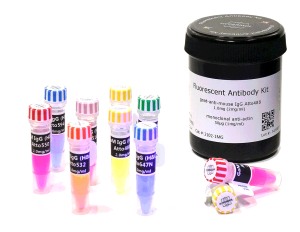


Fluorescent Antibody Kit
Goat Anti-Guinea Pig IgG (H+L) ATTO488
Antibody produced in goat, affinity purified
Cat. # 2703-1MG
............................................................................................................................................................................................................................................................................................................................................................................................................
Description
|
Product name |
Goat Anti-Guinea Pig IgG ATTO488 |
|
Target species |
Guinea Pig |
|
Description |
ATTO-labelled secondary antibodies belong to the new generation of fluorescent antibodies characterized by their exceptional fluorescence intensity and superior photostability. |
|
Specification |
Goat anti-Guinea Pig- IgG ATTO488 (H+L), Immunofluorescence: 1:500-1:1500, Degree of labeling (DOL) : 2-9, Unconjugated dye ≤5% of total fluorescence. |
|
Optical Properties |
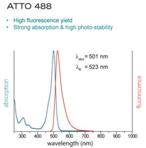
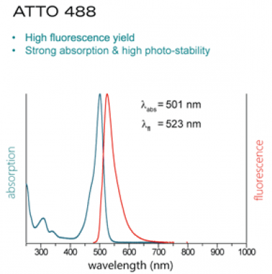
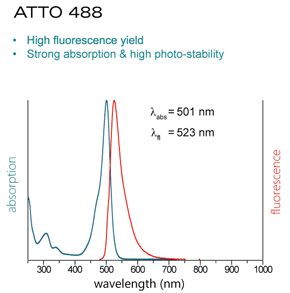
λex 501nm, λem 523nm |
Properties
|
Form |
Lyophilized |
|
Nominal concentration |
2 mg/ml (after reconstitution with buffer) |
|
Content |
1 mg Goat Anti-Guinea Pig- IgG (H+L) ATTO488 |
|
Clonality |
Polyclonal |
|
Isotype |
IgG |
|
Purification notes |
Column affinity purified. |
|
Storage instructions |
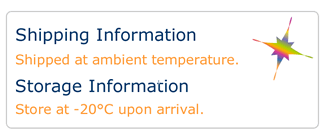
Store as glycerol stock at -20°C. Avoid repeated freeze / thaw cycles. |
|
Shipping conditions |
Shipped at ambient temperature. |
|
Remarks |
For Use in Research only. Not for Use in Human or Veterinary Diagnostical or Therapeutical Applications. |
|
CAS no. |
Further Information
 Product DataSheet
  Material and Safety Data Sheet
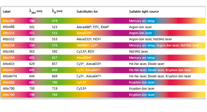 Table of Dyes and Light Sources
 Catalogue: ATTO Secondary Antibodies
References
Super-multiplex vibrational imaging.
Wei L, Chen Z, Shi L, Long R, Anzalone AV, Zhang L, Hu F, Yuste R, Cornish VW, Min W.
Nature. 2017 Apr 27;544(7651):465-470. doi: 10.1038/nature22051. Epub 2017 Apr 19.
αv Integrins combine with LC3 and atg5 to regulate Toll-like receptor signalling in B cells.
Acharya M, Sokolovska A, Tam JM, Conway KL, Stefani C, Raso F, Mukhopadhyay S, Feliu M, Paul E, Savill J, Hynes RO, Xavier RJ, Vyas JM, Stuart LM, Lacy-Hulbert A.
Nat Commun. 2016 Mar 11;7:10917. doi: 10.1038/ncomms10917.
Mechanical signaling coordinates the embryonic heartbeat.
Chiou KK, Rocks JW, Chen CY, Cho S, Merkus KE, Rajaratnam A, Robison P, Tewari M, Vogel K, Majkut SF, Prosser BL, Discher DE, Liu AJ.
Proc Natl Acad Sci U S A. 2016 Aug 9;113(32):8939-44. doi: 10.1073/pnas.1520428113. Epub 2016 Jul 25.
Product DataSheet
Material and Safety Data SheetTable of Dyes and Light Sources Catalogue: ATTO Secondary Antibodies
Wei L, Chen Z, Shi L, Long R, Anzalone AV, Zhang L, Hu F, Yuste R, Cornish VW, Min W.
Nature. 2017 Apr 27;544(7651):465-470. doi: 10.1038/nature22051. Epub 2017 Apr 19.
αv Integrins combine with LC3 and atg5 to regulate Toll-like receptor signalling in B cells.
Acharya M, Sokolovska A, Tam JM, Conway KL, Stefani C, Raso F, Mukhopadhyay S, Feliu M, Paul E, Savill J, Hynes RO, Xavier RJ, Vyas JM, Stuart LM, Lacy-Hulbert A.
Nat Commun. 2016 Mar 11;7:10917. doi: 10.1038/ncomms10917.
Mechanical signaling coordinates the embryonic heartbeat.
Chiou KK, Rocks JW, Chen CY, Cho S, Merkus KE, Rajaratnam A, Robison P, Tewari M, Vogel K, Majkut SF, Prosser BL, Discher DE, Liu AJ.
Proc Natl Acad Sci U S A. 2016 Aug 9;113(32):8939-44. doi: 10.1073/pnas.1520428113. Epub 2016 Jul 25.