

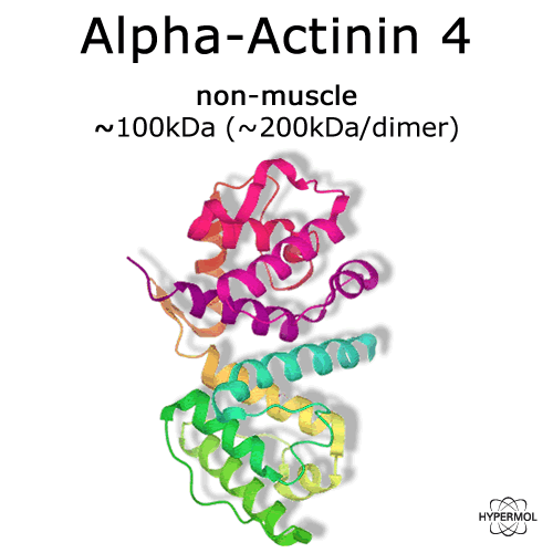
α-Actinin 4
(non-muscle isoform)
Cat. #: 8311-01
.....................................................................................................................................................................................................................................................................................................................................................................................................................................................
Description
|
|
|
|
Protein |
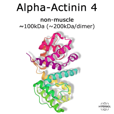
α-Actinin 4 (non-muscle) |
|
Origin |
murine, recombinant |
|
Molecular mass |
~100 kDa each monomer / ~200kDa per dimer |
|
Protein description |
α-Actinins belong to the spectrin superfamily and are divided in four isoforms, encoded by different genes. Isoform 2 and 3 – the so called "muscle" α-actinins are constituents of sarcomeres in the cross-striated skeletal and cardiac muscle. Isoform 1 and 4 are "non-muscle" α-actinins and present in practically every tissue. |
|
Actin interaction |
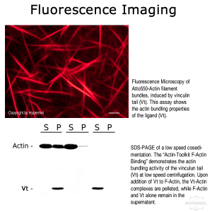
Actin cross linking protein. Kd actin-binding = 1-5µM. |
Properties
|
|
|
|
Form |
Lyophilized, ready-to-use. |
|
Quantity per unit |
2x50 µg |
|
Buffer |
100mM NaCl, 20mM Tris-cl pH 7.4, 0.5mM DTT and 3% disaccharides, when reconstituted with 50µl ultrapure water to obtain a 1mg/ml solution. |
|
Purity |
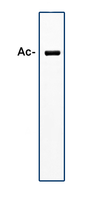
>98% by scanning densitometry from Coomassie G-250 stained SDS-Gels. |
|
Purification notes |
Purified from turkey gizzard muscle (IEX, SEC). |
|
Protein concentration |
Determined UV-VIS (A280=0.1201) |
|
Storage instructions |
Upon reconstitution the product is stored on ice. This product can also be stored as a glycerol stock at -20°C. Avoid repeated freeze / thaw cycles. |
|
Shipping conditions |
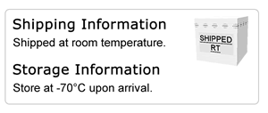
At ambient temperature. Upon delivery store at -70°C. |
|
Remarks |
For Use in Research only. Not for Use in Human or Veterinary Diagnostical or Therapeutical Applications. |
|
CAS no. |
|
Further Information
 Product DataSheet
 Material and Safety Data Sheet
 Protein Sequence on NCBI
References
Real-time dynamics of emerging actin networks in cell-mimicking compartments.
Deshpande S, Pfohl T.; PLoS One. 2015 Mar 18;10(3):e0116521. doi: 10.1371/journal.pone.0116521. eCollection 2015.
A high molecular mass protein isolated from chicken gizzard: its localization at the dense plaques and dense bodies of smooth muscle and the Z-disks of skeletal muscle.
Terasaki AG, Nakagawa H, Kotani E, Mori H, Ohashi K.; J Cell Sci. 1995 Mar;108 ( Pt 3):857-68
Product DataSheetMaterial and Safety Data SheetProtein Sequence on NCBI (chain A)Protein Sequence on NCBI (chain B)Amino acid sequence of
Alpha-actinin-4, mus musculus, untagged
MVDYHAANQAYQYGPNSGGGNGAGGGGSMGDYMAQEDDWDRDLLLDPAWEKQQRKTFTAW
CNSHLRKAGTQIENIDEDFRDGLKLMLLLEVISGERLPKPERGKMRVHKINNVNKALDFI
ASKGVKLVSIGAEEIVDGNAKMTLGMIWTIILRFAIQDISVEETSAKEGLLLWCQRKTAP
YKNVNVQNFHISWKDGLAFNALIHRHRPELIEYDKLRKDDPVTNLNNAFEVAEKYLDIPK
MLDAEDIVNTARPDEKAIMTYVSSFYHAFSGAQKAETAANRICKVLAVNQENEHLMEDYE
RLASDLLEWIRRTIPWLEDRVPQKTIQEMQQKLEDFRDYRRVHKPPKVQEKCQLEINFNT
LQTKLRLSNRPAFMPSEGRMVSDINNGWQHLEQAEKGYEEWLLNEIRRLERLDHLAEKFR
QKASIHEAWTDGKEAMLKQRDYETATLSDIKALIRKHEAFESDLAAHQDRVEQIAAIAQE
LNELDYYDSHNVNTRCQKICDQWDNLGSLTHSRREALEKTEKQLETIDQLHLEYAKRAAP
FNNWMESAMEDLQDMFIVHTIEEIEGLISAHDQFKSTLPDADREREAILAIHKEAQRIAE
SNHIKLSGSNPYTTVTPQIINSKWEKVQQLVPKRDHALLEEQSKQQSNEHLRRQFASQAN
MVGPWIQTKMEEIGRISIEMNGTLEDQLSHLKQYERSIVDYKPSLDLLEQQHQLIQEALI
FDNKHTNYTMEHIRVGWEQLLTTIARTINEVENQILTRDAKGISQEQMQEFRASFNHFDK
DHGGALGPEEFKACLISLGYDVENDRQGDAEFNRIMSVVDPNHSGLVTFQAFIDFMSRET
TDTDTADQVIASFKVLAGDKNFITAEELRRELPPDQAEYCIARMAPYQGPDAAPGALDYK
SFSTALYGESDL
Real-time dynamics of emerging actin networks in cell-mimicking compartments.
Deshpande S, Pfohl T.; PLoS One. 2015 Mar 18;10(3):e0116521. doi: 10.1371/journal.pone.0116521. eCollection 2015.
A high molecular mass protein isolated from chicken gizzard: its localization at the dense plaques and dense bodies of smooth muscle and the Z-disks of skeletal muscle.
Terasaki AG, Nakagawa H, Kotani E, Mori H, Ohashi K.; J Cell Sci. 1995 Mar;108 ( Pt 3):857-68