

Actin Filaments
freshly prepared from rabbit skeletal muscle α-actinCat. #: 8101-F
.............................................................................................................................................................................................................................................................................................................................................................................................................................................................
Description
|
Protein |
Actin (α-Actin) - Actin filaments (F-Actin) |
|
Origin |
Skeletal muscle alpha actin, rabbit |
|
Molecular mass |
42kDa |
|
Protein description |
Freshly prepared actin filaments are shipped on ice by overnight express, ready to start your assays
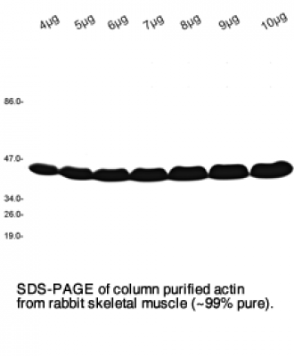
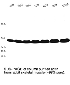
G-Actin prepared from rabbit skeletal muscle is a single chain polypeptide with a molecular weight of 42kD ( 375 amino acids). Actin filaments are obtained by salt induced polymerization of G-Actin. G-Actin from Hypermol is column purified (>99% purity) and pre-centrifuged at 100.000xg, 3h prior to initiation of polymerization. |
|
Purity |
>99% by scanning densitometry. |
Properties
|
Form |
Liquid |
|
Quantity & Concentration |
2 mg ad 2 mg/ml (for other quantities see above). |
|
Buffer |
0.1M KCl, 1.0mM ATP, 2.0mM MgCl2. Buffer: Imidazole pH 7.4. |
|
Availability / Manufacturing time |
2-4 days. |
|
Purification notes |
Purified from rabbit skeletal muscle, GPC. |
|
Protein concentration |
Determined by the Biuret method. |
|
Storage instructions |
F-Actin is kept on ice and should be used within 2-3 weeks. A fresh 100mM ATP solution (is shipped with the product to add 1.0mM after each week of storage to prevent denaturing of actin. Do not freeze. |
|
Shipping conditions |
Wet ice |
|
Remarks |
For Use in Research only. Not for Use in Human or Veterinary Diagnostical or Therapeutical Applications. |
|
CAS no. |
51005-14-2 |
Further Information
 Product DataSheet
 Material and Safety Data Sheet
 Protein Sequence on NCBI
References
Cosolvent and crowding effects on the polymerization kinetics of actin.
Rosin, Christopher, Paul Hendrik Schummel and Robert S. C. Winter.
Physical chemistry chemical physics : PCCP 17 13 (2015): 8330-7.
Exploring the stability limits of actin and its suprastructures.
Rosin C, Erlkamp M, Ecken JV, Raunser S, Winter R.
Biophys J. 2014 Dec 16;107(12):2982-2992. doi: 10.1016/j.bpj.2014.11.006.
Multivalent cross-linking of actin filaments and microtubules through the microtubule-associated protein Tau.
Cabrales Fontela Y, Kadavath H, Biernat J, Riedel D, Mandelkow E, Zweckstetter M.
Nat Commun. 2017 Dec 7;8(1):1981. doi: 10.1038/s41467-017-02230-8.

- A short introduction to actin :
-
A short introduction to actin
Actin is an abundant structural protein (~42 kDa) of eukaryotes and has a conservation of the primary structure of more than 95%. Thus, actin is one of the most highly-conserved proteins [1]. G-actin is the monomeric, globular form of actin and the subunit of actin filaments. G-actin can be described as consisting of a large and a small domain which together form a cleft that binds ATP, ADP*Pi and ADP, as well as and divalent cations (Ca++ and Mg++ are the physiological relevant ions). [2, 3].
Actin monomers assemble to actin filaments in the presence of e.g. mono- and/or divalent cations. This polymerization process of actin filaments, requires ATP bound to G-actin. Addition of ATP-G-actin is favored at the (+)-end of actin filaments. Hydrolysis of ATP to ADP and the steady release of Pi on G-actin cause a conformational change leading to the disassembly of G-actin-GDP at the (-)-end of actin filaments (treadmilling). Actin-binding proteins like profilin and thymosin-β4 control the intracellular pool of G-actin and inhibit spontaneous nucleation of actin filaments.
Models for the actin filament suggest, that when single actin strands form, two asymmetric actin monomers align to form a twofold axis of symmetry [4-9]; their subsequent assembly into a filament that is composed of a pair of strands causes a left-handed helical twist when the adjacent subunits are positioned with respect to each other.
Higher eukaryotes commonly express several actin isoforms by a family of related genes. Actin isoforms are divided into three classes (alpha [α], beta [β] and gamma [γ]) according to their isoelectric point. In general, alpha actins are found in muscle (α-skeletal, α-aortic smooth, α-cardiac, and γ2-enteric smooth), whereas beta and gamma isoforms are prominent in non-muscle cells (β- and γ1-cytoplasmic).
Functions
Actin structures on PDBe
References
1 Elzinga M., Collins JH., Kuehl WM. & Adelstein RS. Complete amino-acid sequence of actin of rabbit skeletal muscle. Proc. Natl. Acad. Sci. U.S.A. 1973; 70(9):2687-91.
2 Suck D., Kabsch W. & Mannherz HG. Three-dimensional structure of the complex of skeletal muscle actin and bovine pancreatic DNAse I at 6-A resolution. Proc. Natl. Acad. Sci. U.S.A. 1981; 78(7):4319-23.
3 Kabsch W., Mannherz HG., Suck D., Pai EF. & Holmes KC. Atomic structure of the actin:DNase I complex. Nature 1990; 347(6288):37-44.
4 Holmes KC., Popp D., Gebhard W. & Kabsch W. Atomic model of the actin filament. Nature 1990; 347(6288):44-9. [PMID: 2395461]
5 Egelman EH. The structure of F-actin. J. Muscle Res. Cell. Motil. 1985; 6(2):129-51.
6 Orlova A., Galkin VE., VanLoock MS., Kim E., Shvetsov A., Reisler E. & Egelman EH. Probing the structure of F-actin: cross-links constrain atomic models and modify actin dynamics. J. Mol. Biol. 2001; 312(1):95-106.
7 Oda T., Stegmann H., Schröder RR., Namba K. & Maéda Y. Modeling of the F-actin structure. Adv. Exp. Med. Biol. 2007; 592:385-401.
8 Aebi U., Fowler WE., Isenberg G., Pollard TD. & Smith PR. Crystalline actin sheets: their structure and polymorphism. J. Cell Biol. 1981; 91(2 Pt 1):340-51.
9 Egelman EH., Francis N. & DeRosier DJ. F-actin is a helix with a random variable twist. Nature 1982; 298(5870):131-5.